


Brain PET scan
Definition
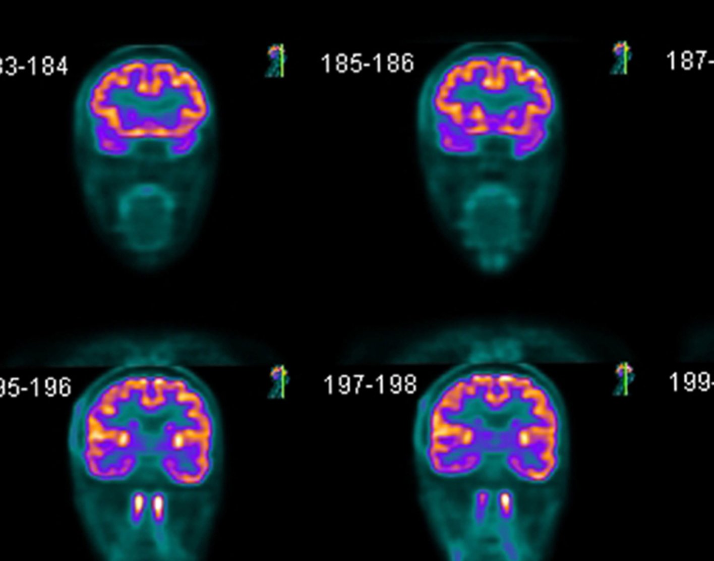
A brain positron emission tomography (PET) scan is an imaging test of the brain. It uses a radioactive substance called a tracer to look for disease or injury in the brain.
A PET scan shows how the brain and its tissues are working. Other imaging tests, such as magnetic resonance imaging (
Alternative Names
Brain positron emission tomography; PET scan - brain
How the Test is Performed
A PET scan requires a small amount of radioactive material (tracer). This tracer is given through a vein (IV), usually on the inside of your elbow. Or, you breathe in the radioactive material as a gas.
The tracer travels through your blood and collects in organs and tissues. The tracer helps your health care provider to see certain areas or diseases more clearly.
You wait nearby as the tracer is absorbed by your body. This usually takes about 1 hour.
Then, you lie on a narrow table, which slides into a large tunnel-shaped scanner. The PET scanner detects signals from the tracer. A computer changes the results into 3-D pictures. The images are displayed on a monitor for your provider to read.
You must lie still during the test so that the machine can produce clear images of your brain. You may be asked to read or name letters if your memory is being tested.
The test takes between 30 minutes and 2 hours.
How to Prepare for the Test
You may be asked not to eat anything for 4 to 6 hours before the scan. You will be able to drink water.
Tell your provider if:
- You are afraid of close spaces (have claustrophobia). You may be given a medicine to help you feel sleepy and less anxious.
- You are pregnant or think you might be pregnant.
- You have any allergies to injected dye (contrast).
- You have taken insulin for diabetes. You will need special preparation.
Always tell your provider about the medicines you are taking, including those bought without a prescription. Sometimes, medicines interfere with the test results.
How the Test will Feel
You may feel a sharp sting when the needle containing the tracer is placed into your vein.
A PET scan causes no pain. The table may be hard or cold, but you can request a blanket or pillow.
An intercom in the room allows you to speak to someone at any time.
There is no recovery time unless you were given a medicine to relax.
After the test, drink a lot of fluids to flush the tracer out of your body.
Why the Test is Performed
A PET scan can show the size, shape, and function of the brain, so your doctor can make sure it is working as well as it should. It is most often used when other tests, such as MRI scan or CT scan, do not provide enough information.
This test can be used to:
- Diagnose cancer
- Prepare for epilepsy surgery
- Help diagnose dementia if other tests and exams do not provide enough information
- Tell the difference between Parkinson disease and other movement disorders
Several PET scans may be taken to determine how well you are responding to treatment for cancer or another illness.
Normal Results
There are no problems detected in the size, shape, or function of the brain. There are no areas in which the tracer has abnormally collected.
What Abnormal Results Mean
Abnormal results may be due to:
Alzheimer disease ordementia due to other health conditionsBrain tumor or spread of cancer from another body area to the brainEpilepsy , and may identify where the seizures start in your brain- Movement disorders (such as
Parkinson disease )
Risks
The amount of radiation used in a PET scan is low. It is about the same amount of radiation as in most CT scans. Also, the radiation does not last for long in your body.
Women who are pregnant or are breastfeeding should let their provider know before having this test. Infants and babies developing in the womb are more sensitive to the effects of radiation because their organs are still growing.
It is possible, though very unlikely, to have an allergic reaction to the radioactive substance. Some people have pain, redness, or swelling at the injection site.
Considerations
It is possible to have false results on a PET scan. Blood sugar or insulin levels may affect the test results in people with
PET scans may be done along with a CT scan. This combination scan is called a PET/CT.
References
Barras CD, Bhattacharya JJ. Current status of imaging of the brain and anatomical features. In: Adam A, Dixon AK, Gillard JH, Schaefer-Prokop CM, eds. Grainger & Allison's Diagnostic Radiology. 7th ed. Philadelphia, PA: Elsevier; 2021:chap 53.
Glaudemans AWJM, Israel O, Ben-Haim S, Slart RHJA. Vascular PET/CT and SPECT/CT. In: Sidawy AN, Perler BA, eds. Rutherford's Vascular Surgery and Endovascular Therapy. 10th ed. Philadelphia, PA: Elsevier; 2023:chap 31.
Jankovic J, Mazziotta JC, Newman NJ, Pomeroy SL. Investigations in the diagnosis and management of neurological disease. In: Jankovic J, Mazziotta JC, Pomeroy SL, Newman NJ, eds. Bradley and Daroff's Neurology in Clinical Practice. 8th ed. Philadelphia, PA: Elsevier; 2022:chap 34.
Review Date: 23/01/2023
The information provided herein should not be used during any medical emergency or for the diagnosis or treatment of any medical condition. A licensed physician should be consulted for diagnosis and treatment of any and all medical conditions. Call 911 for all medical emergencies. Links to other sites are provided for information only -- they do not constitute endorsements of those other sites. Copyright ©2019 A.D.A.M., Inc., as modified by University of California San Francisco. Any duplication or distribution of the information contained herein is strictly prohibited.
Information developed by A.D.A.M., Inc. regarding tests and test results may not directly correspond with information provided by UCSF Health. Please discuss with your doctor any questions or concerns you may have.