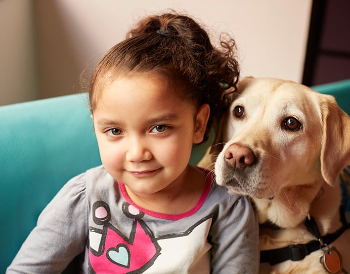
Tender wagging care
Our therapy dogs spread joy and smiles at the bedside and throughout the hospital.
Visit Child Life services

Choledochal cysts are abnormally enlarged areas of bile ducts, the tubes that carry bile from the liver into the small intestine to digest the fats from food. The cysts are benign (noncancerous) at the time of diagnosis.
If bile builds up within choledochal cysts, gallstones can form, which can block the flow of bile through the liver. Without treatment, this blockage can cause cholangitis (infection of the bile ducts), pancreatitis (inflammation of the pancreas) and, in some cases, cirrhosis (severe liver damage). Over the long term, untreated choledochal cysts can lead to gallbladder or bile duct cancer.
Choledochal cysts are congenital, meaning they are present from birth. Symptoms may appear shortly after birth, several years later or not until adulthood. The cause of choledochal cysts is unknown.
This rare condition affects about one in 100,000 children in the United States. Girls are more often diagnosed than boys, as are children of East Asian descent.

Top 10 in the nation for gastroenterology & GI surgery

Ranked among the nation's best in 11 specialties
Choledochal cysts are classified into five categories based on their shape and where they appear in the body. These are:
Choledochal cysts are present from birth, but the first symptoms may appear in infancy, childhood or later in life. These include:
Choledochal cysts can sometimes be seen prenatally during a pregnancy ultrasound and diagnosed before birth. This can allow your child's provider to start planning early for treatment.
For other children, doctors will conduct a physical exam, gather details of medical history, and order blood tests that can help assess bile flow and liver health.
If choledochal cysts are suspected, the next step is to collect images of your child's bile ducts and surrounding organs. This helps your child's doctor find the cyst or cysts and make a plan for treatment. Imaging techniques may include:
If a choledochal cyst is causing a bile duct blockage, treatment involves removing the blockage, then addressing any associated problems, such as infection or injury to the liver or pancreas. A blockage can often be removed using minimally invasive procedures, such as such as endoscopic retrograde cholangiopancreatography (ERCP) or percutaneous transhepatic cholangiography (PTC).
For cysts outside the liver, treatment involves surgically removing each cyst. After the affected part of the bile duct is removed, the intestine is reattached to the remaining bile duct or directly to the liver. If cysts are found inside the liver, the surgeon may need to remove a portion of the organ along with the cysts.
Often, surgeons can remove choledochal cysts through laparoscopy, a form of minimally invasive surgery done through small incisions. Your child's surgeon uses a tiny camera and medical instruments to locate and remove each cyst, which can lead to a faster recovery and less postoperative pain than with conventional surgery.
After surgery, patients still have a small risk for bile duct scarring or narrowing, bile duct infection and bile duct cancer, so it's important to get regular checkups to ensure that the bile ducts are in good condition.
UCSF's providers have extensive experience using minimally invasive surgery to treat choledochal cysts, as well as in monitoring with regular blood work and imaging tests to ensure children recover quickly and stay healthy.
UCSF Benioff Children's Hospitals medical specialists have reviewed this information. It is for educational purposes only and is not intended to replace the advice of your child's doctor or other health care provider. We encourage you to discuss any questions or concerns you may have with your child's provider.
Tender wagging care
